



RNA Mods & Immunology Blog 1



RNA Modifications in Immune Regulation
The immune system responds to infection, tissue damage, and endogenous stress while maintaining tolerance to healthy tissues. Dendritic cells and macrophages initiate inflammatory responses and help activate adaptive immunity, while natural killer (NK) cells eliminate infected or abnormal cells.1 T cells coordinate immune responses and eliminate abnormal cells, while regulatory T cells (Tregs) help maintain self-tolerance.2 B cells produce antibodies and contribute to immune memory.1 Together, these cell populations continuously adjust their activation, differentiation, and function as immune conditions change.
RNA modifications are chemical marks added to RNA after transcription, collectively known as the epitranscriptome, and they provide an important layer of post-transcriptional regulation relevant to the functional immune state. N6-methyladenosine (m6A), one of the most extensively studied RNA modifications, is regulated by writer, eraser, and reader proteins that influence RNA stability, translation, and other aspects of RNA metabolism. A-to-I RNA editing, catalyzed by ADAR enzymes, has a distinct role in controlling endogenous double-stranded RNA (dsRNA) and its recognition by innate immune sensors. Pseudouridine (Ψ), another abundant RNA modification, can also alter innate immune recognition of RNA, with pseudouridine-containing RNA showing reduced activation of Toll-like receptors compared with unmodified RNA.3 These different mechanisms influence immune-cell development, differentiation, activation, and function.4,5
m6A and Immune Cell Function
Emerging evidence indicates that m6A regulates both innate and adaptive immunity, with effects reported across T cells, B cells, dendritic cells, macrophages, and NK cells.4,5 The underlying mechanisms vary by cell type but include regulation of cytokine signaling, antigen presentation, and effector function through changes in the stability and translation of specific target transcripts (Table 1, Figure 1).
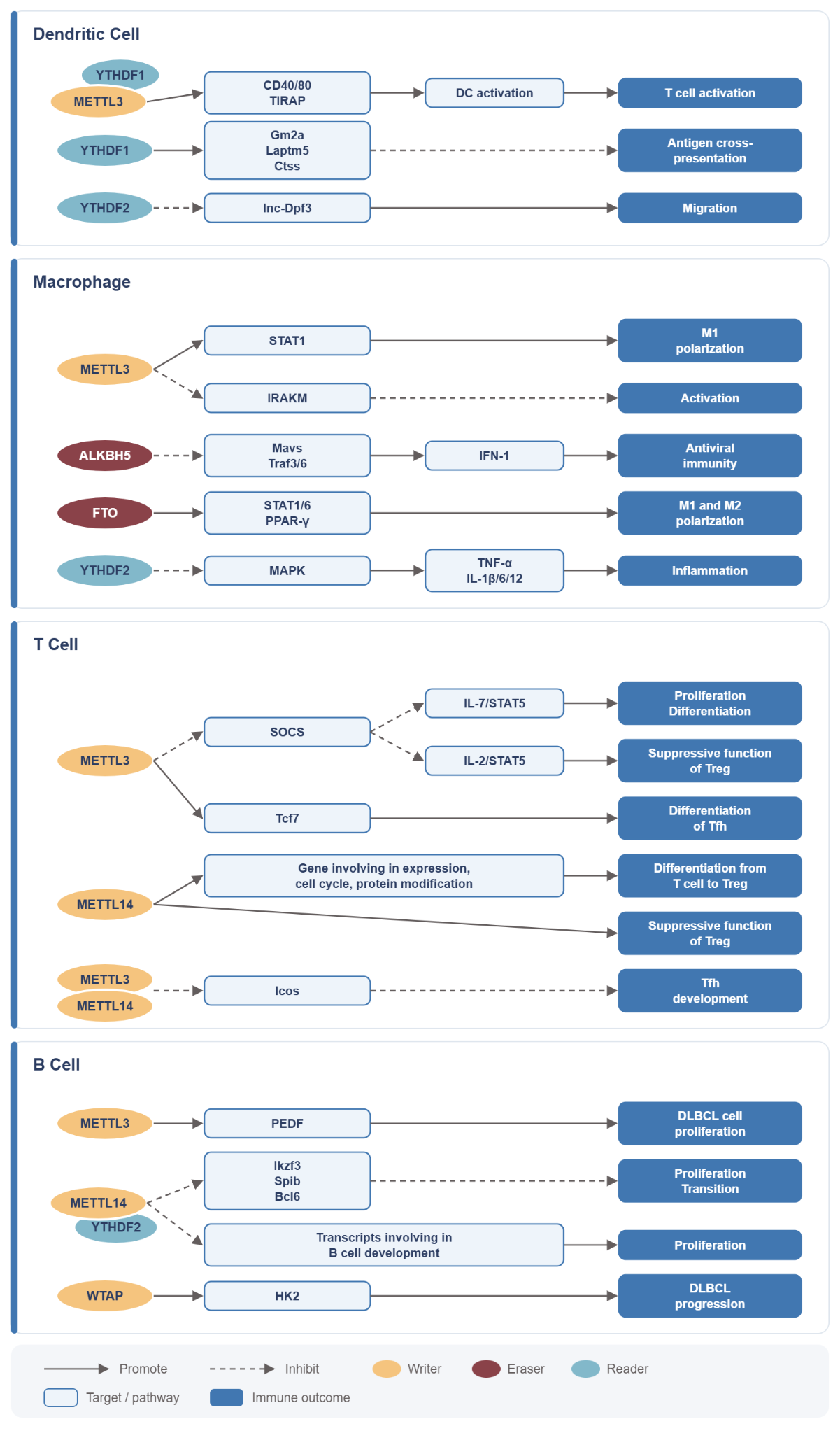
In T cells, m6A destabilizes negative regulators of cytokine signaling, reinforcing pathways that govern naive T-cell homeostasis and differentiation.6 A related mechanism sustains the suppressive function of regulatory T cells (Tregs), linking m6A regulation directly to immune tolerance.7
In dendritic cells, m6A acts at two distinct points. It promotes translation of costimulatory and signaling proteins that drive dendritic-cell maturation,8 while the m6A reader YTHDF1 regulates antigen processing; loss of YTHDF1 increases tumor-antigen cross-presentation and CD8+ T-cell priming.9 m6A has also been implicated in macrophage polarization and NK-cell maturation and effector function, although these mechanisms are less thoroughly characterized.4,5
m6A and Autoimmune Disease
Disruption of m6A regulation has increasingly been linked to autoimmune and inflammatory disease through several distinct mechanisms. The examples below illustrate how altered m6A regulation can affect different cell types and pathways across autoimmune diseases.
Sjögren’s disease. Sjögren’s is a chronic autoimmune disease that primarily affects the salivary and lacrimal glands, causing dry mouth and eyes, although other organs can also be affected. Glandular dysfunction is accompanied by immune activation of epithelial cells and infiltration by T and B cells.10 Recent work identified an epitranscriptomic mechanism connecting m6A directly with endogenous RNA surveillance in glandular epithelial cells. METTL3-dependent m6A stabilizes the mRNA encoding REXO2, a mitochondrial exonuclease involved in clearing mitochondrial dsRNA.11 When this pathway is disrupted, REXO2 expression declines, mitochondrial dsRNA accumulates, and RNA-sensing pathways drive type I interferon signaling. Reduced REXO2 was also observed in salivary-gland epithelium from patients with Sjögren’s disease, linking the experimentally defined pathway with human autoimmunity.
Systemic lupus erythematosus. SLE is a systemic autoimmune disease that predominantly affects women and can involve the skin, joints, kidneys, and other organs. Its heterogeneous immune dysfunction includes autoreactive B cells and plasma cells that produce autoantibodies, together with dysregulated T-cell and interferon responses.12 A distinct m6A mechanism has been described in SLE, where METTL3 promotes m6A-dependent IRF4 expression, associated with increased plasma-cell infiltration and kidney injury in lupus nephritis.13 This connects altered m6A regulation with a B-cell lineage central to SLE pathology.
Multiple sclerosis. MS is a major chronic immune-mediated disease of the central nervous system, particularly affecting young adults, and is characterized by demyelination and neuroaxonal injury. T cells, B cells, and myeloid cells contribute to CNS inflammation and tissue damage.14 In experimental autoimmune encephalomyelitis (EAE), a widely used animal model of MS, T-cell-specific loss of the m6A demethylase ALKBH5 increased m6A modification of Ifng and Cxcl2 transcripts. In this context, increased methylation accelerated transcript decay, reducing IFN-γ and CXCL2 expression, pathogenic CD4+ T-cell responses, neutrophil recruitment, and disease severity.15 This illustrates an important feature of m6A biology: the functional consequence of methylation is transcript- and context-dependent rather than uniformly stabilizing or destabilizing RNA. Because these experiments were performed in an animal model, the extent to which this mechanism operates in human MS remains to be established.
m6A can also regulate tissue responses that contribute to inflammatory disease. In the intestinal epithelium, m6A methylation regulates Nfkbia mRNA stability and NF-κB-mediated epithelial survival and homeostasis, providing another connection between RNA modification and inflammatory signaling.16 Together, these studies show that epitranscriptomic regulation can influence disease through immune-cell differentiation, epithelial homeostasis, inflammatory signaling, and endogenous RNA surveillance.
A-to-I RNA Editing and Self-RNA Recognition
A-to-I editing provides a mechanistically distinct layer of protection against inappropriate immune activation. ADAR1 edits endogenous double-stranded RNA (dsRNA), limiting its recognition by innate immune sensors. When ADAR1-mediated editing is insufficient, endogenous dsRNA can activate MDA5 and downstream MAVS signaling, producing a type I interferon response even in the absence of infection.17
This mechanism also contributes directly to immune tolerance. In mice, T-cell-specific loss of ADAR1 disrupts thymocyte maturation and negative selection, increases interferon-stimulated gene expression, and produces autoimmune pathology including spontaneous colitis. Concurrent deletion of MDA5 rescues thymocyte maturation and prevents disease, demonstrating that ADAR1-mediated editing of endogenous RNA helps prevent inappropriate MDA5 activation during establishment of T-cell self-tolerance.18
Human genetic studies further connect RNA-editing variation with susceptibility to common inflammatory diseases, including SLE (Systemic lupus erythematosus), RA (Rheumatoid Arthritis), MS (Multiple Sclerosis), and IBD (Inflammatory Bowel Disease), and identify disease-associated loci at which genetically reduced editing is linked to inflammatory signaling.19 These observations extend the experimentally established ADAR1-MDA5 mechanism toward human inflammatory disease, although the contribution of specific editing sites and cell types remains an active area of investigation.
Considered alongside the mechanism shown for Sjögren’s disease, these findings reveal a broader principle. m6A-dependent stabilization of REXO2 helps prevent mitochondrial dsRNA accumulation, whereas A-to-I editing modifies endogenous dsRNA to prevent inappropriate recognition by MDA5. Different RNA-modification pathways can therefore converge on a common function: preventing the cell’s own RNA from becoming an inflammatory signal.
Toward Epitranscriptomic Biomarkers of Immune State
Current approaches to monitoring autoimmune disease, including clinical activity scores, autoantibody testing, inflammatory protein panels, and transcriptomic profiling, largely capture the downstream consequences of immune dysregulation rather than the underlying regulatory processes that shape immune state. The mechanisms described above suggest that RNA modification state may provide additional information not captured by transcript abundance alone.
Longitudinal blood-based RNA profiling has already demonstrated a version of this predictive capability in RA. In patients with RA, repeated home blood collection over multiple years enabled longitudinal RNA sequencing that identified a transcriptional signature arising from a cell population the authors termed PRIME cells. These cells appeared in blood one to two weeks before clinical flares, ahead of joint symptom onset.20 This result relied on conventional transcript-level sequencing rather than RNA-modification profiling, but it establishes that longitudinal blood RNA measurement can anticipate autoimmune flares before they become clinically apparent, raising the question of whether modification state could provide complementary or earlier signals within a similar sampling strategy.
A separate line of correlative evidence comes from SLE directly. A cross-sectional study of PBMCs from SLE patients found that mRNA levels of the m6A writer METTL14, eraser ALKBH5, and reader YTHDF2 were reduced compared with healthy controls and that these reductions correlated with several clinical and laboratory markers of disease activity.21 This study measured expression of the m6A regulatory machinery itself rather than modification state on specific transcripts, and the comparison was made at a single timepoint rather than across a patient’s own disease course.
Together, these findings motivate further investigation into whether RNA modification profiling in longitudinal blood sampling could provide additional or earlier information about immune activation in autoimmune disease. Whether such modification signatures can serve as biomarkers of immune tolerance or disease activity remains an important question for prospective, longitudinal studies designed for that purpose.
Conclusion
RNA modifications are highly impactful on immune regulation. They operate across innate and adaptive immune cells, influencing their development, differentiation, activation, and function. m6A regulates processes ranging from T-cell differentiation and immune tolerance to dendritic-cell maturation and antigen presentation, while m6A-dependent mitochondrial RNA surveillance and A-to-I editing help prevent endogenous RNA from inducing inappropriate inflammatory responses.
As profiling technologies mature, measuring RNA modifications alongside gene expression offers an opportunity to investigate both how genes are expressed and how their RNAs are functionally regulated. This additional information has strong promise to improve our understanding of immune state, disease activity, and therapeutic response.
Explore RNA modifications in your research
EpiPlex® enables scalable profiling of m6A, inosine, pseudouridine, and gene expression across a range of research and clinical sample types, with integrated bioinformatics for analysis and interpretation. Learn more about EpiPlex or talk with us about your research.
Explore additional posts in our Mods in Motion series
Blog Posts
- The Power of Inosine: How RNA Editing Shapes the Transcriptome
- A Beginner’s Guide to m6A and Other RNA Modifications
- Beyond Transcriptomics: RNA Modifications Shape Cancer Progression
Podcasts
About the author
James Tsay
James M. Tsay, PhD, is a biotech scientist and industry leader whose work spans across genomics, molecular diagnostics, and multi-omic technologies. He earned his PhD in physical chemistry at UCLA and completed postdoctoral research in biophysics at UC San Diego. He is currently a Scientific Fellow at AlidaBio, and previously held senior scientific and technical leadership roles at Illumina and Pleno Biosciences, contributing to the development of next-generation sequencing and advanced molecular profiling platforms.
References
- Iwasaki, A., Medzhitov, R. (2015). Control of adaptive immunity by the innate immune system. Nature Immunology, 16(4), 343-353. https://doi.org/10.1038/ni.3123
- Sakaguchi, S., Yamaguchi, T., Nomura, T., Ono, M. (2008). Regulatory T cells and immune tolerance. Cell, 133(5), 775-787. https://doi.org/10.1016/j.cell.2008.05.009
- Bérouti, M., Wagner, M., Greulich, W., et al. (2025). Pseudouridine RNA avoids immune detection through impaired endolysosomal processing and TLR engagement. Cell, 188(18), 4880-4895.e15. https://doi.org/10.1016/j.cell.2025.05.032
- Cui, L., Ma, R., Cai, J., et al. (2022). RNA modifications: importance in immune cell biology and related diseases. Signal Transduction and Targeted Therapy, 7, 334. https://doi.org/10.1038/s41392-022-01175-9
- Wang, Y., Li, L., Li, J., Zhao, B., Huang, G., Li, X., Xie, Z., Zhou, Z. (2021). The emerging role of m6A modification in regulating the immune system and autoimmune diseases. Frontiers in Cell and Developmental Biology, 9, 755691. https://doi.org/10.3389/fcell.2021.755691
- Li, H. B., Tong, J., Zhu, S., et al. (2017). m6A mRNA methylation controls T cell homeostasis by targeting the IL-7/STAT5/SOCS pathways. Nature, 548(7667), 338-342. https://doi.org/10.1038/nature23450
- Tong, J., Cao, G., Zhang, T., et al. (2018). m6A mRNA methylation sustains Treg suppressive functions. Cell Research, 28(2), 253-256. https://doi.org/10.1038/cr.2018.7
- Wang, H., Hu, X., Huang, M., et al. (2019). Mettl3-mediated mRNA m6A methylation promotes dendritic cell activation. Nature Communications, 10, 1898. https://doi.org/10.1038/s41467-019-09903-6
- Han, D., Liu, J., Chen, C., et al. (2019). Anti-tumour immunity controlled through mRNA m6A methylation and YTHDF1 in dendritic cells. Nature, 566, 270-274. https://doi.org/10.1038/s41586-019-0916-x
- Baldini, C., Fulvio, G., La Rocca, G., et al. (2024). Update on the pathophysiology and treatment of primary Sjögren syndrome. Nature Reviews Rheumatology, 20, 473-491. https://doi.org/10.1038/s41584-024-01135-3
- Arco-Hierves, A., Pamboukas, K., Bilonda Mutala, L., et al. (2026). Loss of epitranscriptomic mitochondrial RNA surveillance drives epithelial type I interferon and inflammation in autoimmunity. Proceedings of the National Academy of Sciences, 123(35), e2534325123. https://doi.org/10.1073/pnas.2534325123
- Caielli, S., Wan, Z., Pascual, V. (2023). Systemic lupus erythematosus pathogenesis: interferon and beyond. Annual Review of Immunology, 41, 533-560. https://doi.org/10.1146/annurev-immunol-101921-042422
- Liu, Y., Wang, X., Huang, M., et al. (2024). METTL3 facilitates kidney injury through promoting IRF4-mediated plasma cell infiltration via an m6A-dependent manner in systemic lupus erythematosus. BMC Medicine, 22, 511. https://doi.org/10.1186/s12916-024-03735-y
- Filippi, M., Bar-Or, A., Piehl, F., et al. (2018). Multiple sclerosis. Nature Reviews Disease Primers, 4, 43. https://doi.org/10.1038/s41572-018-0041-4
- Zhou, J., Zhang, X., Hu, J., et al. (2021). m6A demethylase ALKBH5 controls CD4+ T cell pathogenicity and promotes autoimmunity. Science Advances, 7(25), eabg0470. https://doi.org/10.1126/sciadv.abg0470
- Zhang, T., Ding, C., Chen, H., et al. (2022). m6A mRNA modification maintains colonic epithelial cell homeostasis via NF-κB-mediated antiapoptotic pathway. Science Advances, 8(12), eabl5723. https://doi.org/10.1126/sciadv.abl5723
- Liddicoat, B. J., Piskol, R., Chalk, A. M., et al. (2015). RNA editing by ADAR1 prevents MDA5 sensing of endogenous dsRNA as nonself. Science, 349(6252), 1115-1120. https://doi.org/10.1126/science.aac7049
- Nakahama, T., Kato, Y., Kim, J. I., et al. (2018). ADAR1-mediated RNA editing is required for thymic self-tolerance and inhibition of autoimmunity. EMBO Reports, 19(12), e46303. https://doi.org/10.15252/embr.201846303
- Li, Q., Gloudemans, M. J., Geisinger, J. M., et al. (2022). RNA editing underlies genetic risk of common inflammatory diseases. Nature, 608(7923), 569-577. https://doi.org/10.1038/s41586-022-05052-x
- Orange, D. E., Yao, V., Sawicka, K., et al. (2020). RNA identification of PRIME cells predicting rheumatoid arthritis flares. New England Journal of Medicine, 383(3), 218-228. https://doi.org/10.1056/NEJMoa2004114
- Luo, Q., Rao, J., Zhang, L., et al. (2020). The study of METTL14, ALKBH5, and YTHDF2 in peripheral blood mononuclear cells from systemic lupus erythematosus. Molecular Genetics & Genomic Medicine, 8(9), e1298. https://doi.org/10.1002/mgg3.1298